Proteins are long chains of amino acids joined by peptide bonds. They carry out most of the chemical work of a cell and also build much of its structure[1]. At the start of the 20th century Emil Fischer and Franz Hofmeister proposed that proteins are linear chains of amino acids, which Fischer called polypeptides[1]. Proteins are made mainly of carbon, hydrogen, oxygen, nitrogen and sulfur; the sulfur comes from the amino acids cysteine and methionine[2].
Most proteins work only after the chain has folded into a particular three-dimensional shape, and that shape is set by the order of the amino acids. Much of what follows comes back to this idea: how the chain is made and folds, what happens when the fold is lost or goes wrong, and how chemists have learned to build whole proteins. The last part turns to protein in food, where digestion takes the chain apart again.
| Building blocks | 20 standard amino acids, used unevenly: alanine and leucine make up about 9% of residues each, cysteine and tryptophan about 1% |
|---|---|
| Typical size | About 300 amino acids per chain |
| Protein or peptide | IUPAC: polypeptides heavier than about 10,000 Da are proteins, and the limit is not precise |
| How many | Some eukaryotic cells have more than 20,000 protein-coding genes, and over 100,000 distinct protein molecules once splicing and modifications are counted |
Peptide, polypeptide or protein
IUPAC defines proteins as natural or synthetic polypeptides with a molecular weight above about 10,000, and adds that the limit is not precise[3]. That corresponds to roughly 90 amino acids. In everyday use the line is drawn lower, often at about 50 residues, below which chains are called peptides, and some molecules sit on it: insulin, with 51 residues in two chains, is called both a peptide hormone and a protein. A protein may consist of one polypeptide chain or of several held together, and some also carry non-amino-acid parts such as metal ions or haem.
The mass, formula and isoelectric point of a sequence can be estimated with the peptide calculator. For a whole protein the mass is the sum of its residues plus one water, and modifications add their own mass[2].
Four broad types
Proteins can be sorted roughly into four groups that differ in composition and behaviour[2]:
- Globular proteins are compact and usually soluble in water. Most enzymes belong here.
- Membrane proteins sit in cell membranes and have more hydrophobic residues, especially in the stretches that cross the membrane.
- Fibrous proteins are long and tough and often built from repeats. Collagen, for example, is built from repeats of Gly-X-Y, in which X is often proline and Y often hydroxyproline (Hyp), a modified proline[2]. Keratin is another fibrous protein.
- Intrinsically disordered proteins have no single fixed shape on their own and exist as a shifting mix of forms; many take on a definite structure only when they bind a partner. They are rich in glycine, serine and proline, and an estimated 20–30% of eukaryotic proteins fall into this group.
One protein can combine several of these: a soluble domain attached to a membrane domain, or a folded domain with a disordered tail[2].
Charge and solubility
Proteins carry acidic side chains (aspartate, glutamate) and basic ones (lysine, arginine, histidine), so their net charge depends on pH. At the isoelectric point (pI) the net charge is zero, so the molecules repel each other least and solubility is usually lowest. Proteins with different pI values can therefore be separated from one another[2]. The range is wide: ornithine decarboxylase has a pI of about 4, aldolase about 10. Across all the proteins of an organism the pI values fall into two clusters, with relatively few proteins near pH 7.4, the pH of most compartments inside the cell[2].
Solubility also depends on what the surface carries. Proteins with many charged and polar residues facing the water dissolve well, while membrane proteins, whose surface is largely hydrophobic, are usually insoluble unless a detergent is added[2]. The pI can be estimated from the sequence with the peptide calculator, but an exact value has to be measured[2].
What proteins do
| Role | What it means | Example |
|---|---|---|
| Enzymes | Speed up chemical reactions | Phenylalanine hydroxylase |
| Structure | Give strength to cells and tissues | Collagen, keratin, actin |
| Movement | Make muscles contract | Myosin |
| Transport and storage | Carry or hold small molecules and ions | Haemoglobin (oxygen), ferritin (iron) |
| Signalling | Carry messages between cells | Growth hormone, insulin |
| Defence | Recognise and fight pathogens | Antibodies such as immunoglobulin G |
Defence proteins are not limited to animals. Plants make thionins, defensins, lectins, protease inhibitors and ribosome-inactivating proteins against insects, fungi, bacteria and viruses[4]. Many of the smaller ones are peptides rather than proteins; see antimicrobial peptides.
How cells make proteins
Whatever its job, every protein starts as a chain assembled by a ribosome, which reads a messenger RNA copy of a gene three nucleotides at a time. Four nucleotides give 4 × 4 × 4 = 64 three-letter codons: 61 of them specify the 20 amino acids, and three (UAA, UAG and UGA) mark the end of the chain. The chain grows from its N-terminus to its C-terminus. A ribosome in a eukaryotic cell, such as a human one, adds about 2 amino acids per second, a bacterial ribosome about 20[5].
The new chain is often changed further. Pieces can be cut off, chemical groups attached and disulfide bonds formed; together with alternative splicing of the RNA, this is why a cell holds far more distinct protein molecules than it has genes[1]. Cutting is also how many peptides are born. Insulin is first made as a single chain, proinsulin. After its three disulfide bonds have formed, a segment is removed from the middle, leaving the two chains of the active hormone. The digestive enzyme chymotrypsin is made the same way from a single-chain precursor, chymotrypsinogen[6].
Four levels of protein structure
Once made, the chain folds, and its structure is usually described at four levels.
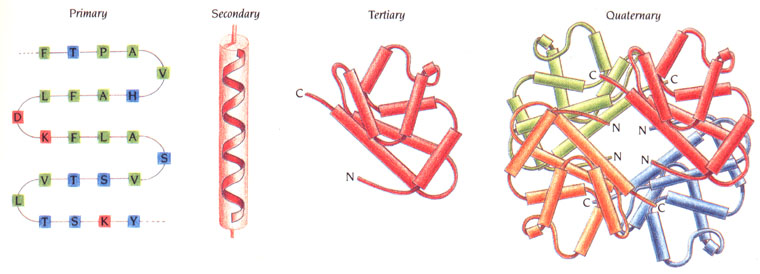
Primary structure is the sequence of amino acids, written from the amino end (N-terminus) to the carboxyl end (C-terminus). It decides how the chain folds, and so what the protein can do[2].
Secondary structure is the regular local folding of the backbone. The two main forms, the α-helix and the β-sheet, were proposed by Linus Pauling, Robert Corey and Herman Branson in 1951[7]. Both are held together by hydrogen bonds between the C=O group of one peptide bond and the N–H group of another. In a helix these bonds run along the axis of a single coil. In a sheet they link neighbouring strands, which can run in the same direction (parallel) or in opposite directions (antiparallel).
Tertiary structure is the overall three-dimensional shape of one chain. It is held by interactions between side chains: hydrophobic side chains gather away from water, and hydrogen bonds, ionic bonds and covalent disulfide bonds lock the fold in place.
Quaternary structure is the assembly of several chains, or subunits, into one working complex[2]. Haemoglobin, for example, is built from four chains.
The sequence decides the shape
Christian Anfinsen showed why the primary structure matters so much. He worked with ribonuclease, an enzyme of 124 amino acids held together by four disulfide bonds. In 8 M urea with a reducing agent the disulfide bonds break and the chain unfolds completely. Its eight cysteines can then pair in 105 different ways, yet when the urea and reducing agent are removed the protein folds back into its active form, with the one correct set of disulfide bonds. If the cysteines are instead allowed to pair while the urea is still present, a mixture of wrongly bonded forms appears, with about 1% of normal activity. A trace of a thiol reagent lets the bonds swap, and the mixture slowly converts into native enzyme[6].
From such experiments Anfinsen concluded that the native shape of a protein is the one of lowest free energy in its normal surroundings, and that it is therefore set by the amino acid sequence. He called this the thermodynamic hypothesis, and it earned him a share of the 1972 Nobel Prize in Chemistry with Stanford Moore and William Stein[6]. Refolding in the test tube took hours, while a cell makes ribonuclease in about two minutes; the gap led to the discovery of an enzyme in the endoplasmic reticulum that speeds up the reshuffling of disulfide bonds[6]. The hypothesis describes proteins with one stable fold. Disordered proteins have no such fold, and in the cell many chains are helped to fold by chaperones[10].
How structures are determined
Knowing that the sequence sets the shape is not the same as knowing the shape. About 90% of the protein structures in the Protein Data Bank were solved by X-ray crystallography, which first requires the protein to be crystallised[2]. Experimental structures are known for around 100,000 unique proteins. That is a small fraction of the billions of known sequences, because a single structure can take months or years of work[8].
Predicting the shape from the sequence alone, the step Anfinsen's work showed to be possible in principle, remained an open problem for more than 50 years. In the blind CASP14 test in 2020, AlphaFold, a neural network developed at DeepMind, predicted structures with a median backbone deviation of 0.96 Å from the experimental ones, measured over 95% of the residues; the next best method managed 2.8 Å. For comparison, a carbon atom is about 1.4 Å wide[8]. Demis Hassabis and John Jumper received half of the 2024 Nobel Prize in Chemistry for this work; the other half went to David Baker for computational protein design[9].
Denaturation
The fold that Anfinsen's ribonuclease found so reliably can also be lost. Denaturation is the loss of a protein's native three-dimensional structure. The peptide bonds stay intact, so the sequence is unchanged; what is lost is the folding held by weaker interactions and, if they are broken too, the disulfide bonds. Heat, extremes of pH, high concentrations of urea or guanidine hydrochloride, and detergents all denature proteins. A denatured protein usually loses its activity.
Whether denaturation can be undone depends on the protein and the conditions. In the kitchen it looks permanent: a boiled egg white stays white when it cools. Yet reversible denaturation was described as early as the 1930s, and small proteins in dilute solution often refold completely. Ribonuclease recovers after full unfolding, and staphylococcal nuclease, a protein of 149 amino acids without disulfide bonds, refolds within seconds[6].
Misfolding and amyloid
Losing the native shape is one problem; taking on a wrong one is another. Peptides and proteins have an inherent tendency to leave their native shape and clump together into amyloid: long, highly ordered fibrils with a cross-β structure, built from stacked β-strands[10]. By 2017, 37 peptides and proteins were known to form amyloid deposits in human disease[10]:
| Peptide or protein | Residues | Disease |
|---|---|---|
| Amyloid-β | 40 or 42 | Alzheimer disease |
| α-Synuclein | 140 | Parkinson disease |
| Islet amyloid polypeptide | 37 | Type II diabetes |
| Prion protein | 208 | Creutzfeldt–Jakob disease |
| Insulin | 30 + 21 | Deposits at injection sites |
| Enfuvirtide | 36 | Deposits at injection sites |
The last two are medicines: even peptide drugs can form amyloid where they are repeatedly injected. Under suitable conditions in the test tube, proteins with no link to any disease, chains made of a single amino acid such as polylysine, and even the free amino acid phenylalanine form fibrils of the same kind[10]. Amyloid is therefore not a peculiarity of a few disease proteins: under suitable conditions, very different chains can adopt it. Cells guard against it with a network of chaperones, which help chains fold, and degradation systems that remove damaged proteins; disease follows when this network is overwhelmed[10].
Making proteins chemically
Most proteins used in research and medicine are produced by cells, but they can also be made entirely by chemistry. Insulin and ribonuclease were synthesised in active form by the classical methods of peptide chemistry[1]. Ribonuclease was also among the first proteins made by solid-phase synthesis, by Bernd Gutte and Bruce Merrifield[6]. Even with the best solid-phase methods, though, small losses at each step added up, and about 50 amino acids remained the longest chain that could be made reliably in one piece[1].
Longer proteins are built from pieces. In native chemical ligation, introduced by Philip Dawson, Stephen Kent and co-workers in 1994, two unprotected peptides react in water at neutral pH and join through a normal peptide bond, in nearly quantitative yield. The full-length chain is then folded into the working protein[1]. Chemical synthesis can do things a cell cannot. It can place any non-standard amino acid or label exactly where it is wanted, and it can build a protein entirely from D-amino acids. Kent's group made both the natural enzyme HIV-1 protease and its mirror image, whose crystal structure proved to be the exact reflection of the natural one[1].
Protein in the diet
Protein is the main structural material of muscle and other tissues, and the body also uses it to make hormones, enzymes and haemoglobin. It can be burned for energy, but it is not the body's preferred fuel[11]. Food proteins are broken down in the gut into amino acids and short peptides, which the body uses to build its own proteins. The recommended dietary allowance for adults is 0.8 g of protein per kilogram of body weight per day[11]. Athletes are generally advised to eat more: about 1.2–1.4 g/kg for endurance athletes and 1.4–1.8 g/kg for strength and power athletes[11]. In one study of strength training, four weeks on 3.3 g/kg a day gave greater gains in protein synthesis and body mass than 1.3 g/kg[11].
How much protein common foods contain, in grams per 100 g of the food as eaten[12]:
| Food | Protein, g |
|---|---|
| Chicken breast, roasted, meat only | 31.0 |
| Beef, ground, 90% lean, broiled | 26.1 |
| Cheddar cheese | 22.9 |
| Peanut butter, smooth | 22.2 |
| Salmon, Atlantic, farmed, cooked | 22.1 |
| Almonds | 21.2 |
| Egg, hard-boiled | 12.6 |
| Greek yoghurt, plain, nonfat | 10.2 |
| Tofu, firm | 9.0–17.3 |
| Lentils, boiled | 9.0 |
| Chickpeas, boiled | 8.9 |
| Black beans, boiled | 8.9 |
| Quinoa, cooked | 4.4 |
| Milk, whole | 3.2 |
Data: USDA National Nutrient Database for Standard Reference, Release 28 (now part of FoodData Central as SR Legacy). Tofu varies with the type: 9.0 g for firm tofu made with calcium sulfate and nigari, 17.3 g for raw firm tofu made with calcium sulfate.
The quality of a protein depends on its content of essential amino acids, which the body cannot make, and on how well it is digested. Animal proteins contain all the essential amino acids and are called complete. Most plant proteins contain too little of one or two of them, so a plant-based diet needs a variety of foods, such as grains and legumes, to supply all of them[11]. The most widely used measure of quality is the protein digestibility corrected amino acid score (PDCAAS), where 1.00 is the top value:
| Protein | PDCAAS |
|---|---|
| Casein, egg, milk, whey | 1.00 |
| Soy protein | 1.00 |
| Beef | 0.92 |
| Black beans | 0.75 |
| Peanuts | 0.52 |
| Wheat gluten | 0.25 |
From Hoffman and Falvo[11], adapted from the U.S. Dairy Export Council (1999) and Sarwar (1997).
PDCAAS has known limits. It may overestimate protein quality in older people, and it is based on digestibility measured over the whole gut rather than at the end of the small intestine, where amino acids are actually absorbed[11]. In a study of bodybuilders eating 2.8 g/kg a day, kidney function tests showed no harmful changes. People who already have kidney disease, however, are often advised to limit their protein intake, which is thought to slow the progression of the disease[11]; the amount is set individually.
Protein supplements
The protein powders used in sports nutrition come mostly from milk and soy. Most of the protein in cow's milk is casein, 70–80% of the total, and about 20% is whey[11]. Whey is sold in three main forms that differ in purity:
| Form | Protein, % | Lactose, % | Fat, % |
|---|---|---|---|
| Whey powder | 11–14.5 | 63–75 | 1–1.5 |
| Whey concentrate | 25–89 | 10–55 | 2–10 |
| Whey isolate | 90+ | 0.5 | 0.5 |
From Hoffman and Falvo[11], adapted from Geiser (2003).
Whey and casein behave differently after a meal. Whey is absorbed quickly and gives a sharp, short rise in amino acids in the blood. Casein forms a clot in the stomach and releases amino acids slowly, sometimes for several hours. In the studies reviewed by Hoffman and Falvo, whey stimulated rapid protein synthesis but much of it was burned as fuel, while casein gave a greater gain of body protein over a longer time[11]. Soy protein is also complete and scores 1.00 on PDCAAS. Its isoflavones have been studied for effects on heart health, with conflicting results so far[11].
This section is general information, not dietary or medical advice.
In short
- A protein is a polypeptide chain, or several, usually heavier than about 10,000 Da; where the peptides end and the proteins begin is settled by convention.
- The amino acid sequence determines how a protein folds. Anfinsen's ribonuclease found its one correct fold among 105 possible disulfide pairings on its own.
- Shape is held mostly by weak interactions, so heat, pH or urea can undo it without breaking a single peptide bond. When folding goes wrong, chains can stack into amyloid, as in Alzheimer and Parkinson disease.
- Many peptides are cut out of larger proteins, insulin from proinsulin being the classic case, and chemistry can now build whole proteins, even in mirror-image form.
- In the diet, adults need about 0.8 g of protein per kg of body weight a day; quality depends on essential amino acids and digestibility.
References
- Kent SBH (2009). Total chemical synthesis of proteins. Chemical Society Reviews 38:338–351. DOI: 10.1039/b700141j
- Tomii K (2019). Protein properties. In: Encyclopedia of Bioinformatics and Computational Biology. Elsevier. DOI: 10.1016/B978-0-12-809633-8.20266-5
- Moss GP, Smith PAS, Tavernier D (1995). Glossary of class names of organic compounds and reactivity intermediates based on structure (IUPAC Recommendations 1995). Pure and Applied Chemistry 67:1307–1375, entry “proteins”. DOI: 10.1351/pac199567081307
- Jain M, Amera GM, Muthukumaran J, Singh AK (2022). Insights into biological role of plant defense proteins: a review. Biocatalysis and Agricultural Biotechnology 40:102293. DOI: 10.1016/j.bcab.2022.102293
- Alberts B, Johnson A, Lewis J, et al. (2002). Molecular Biology of the Cell, 4th edition, chapter “From RNA to protein”. New York: Garland Science. NCBI Bookshelf NBK26829
- Anfinsen CB (1973). Principles that govern the folding of protein chains. Science 181:223–230. DOI: 10.1126/science.181.4096.223
- Pauling L, Corey RB, Branson HR (1951). The structure of proteins: two hydrogen-bonded helical configurations of the polypeptide chain. Proceedings of the National Academy of Sciences USA 37:205–211. DOI: 10.1073/pnas.37.4.205
- Jumper J, Evans R, Pritzel A, et al. (2021). Highly accurate protein structure prediction with AlphaFold. Nature 596:583–589. DOI: 10.1038/s41586-021-03819-2
- The Nobel Prize in Chemistry 2024. NobelPrize.org. nobelprize.org
- Chiti F, Dobson CM (2017). Protein misfolding, amyloid formation, and human disease: a summary of progress over the last decade. Annual Review of Biochemistry 86:27–68. DOI: 10.1146/annurev-biochem-061516-045115
- Hoffman JR, Falvo MJ (2004). Protein – which is best? Journal of Sports Science and Medicine 3:118–130. PMC3905294
- US Department of Agriculture, Agricultural Research Service, Nutrient Data Laboratory (2016). USDA National Nutrient Database for Standard Reference, Release 28. Now available in FoodData Central (SR Legacy). Entries 05064, 23563, 01009, 16398, 15237, 12061, 01129, 01256, 16126, 16426, 16070, 16057, 16015, 20137, 01077.